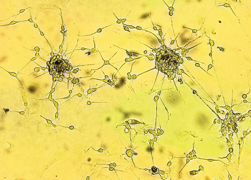
Laboratory experiments reveal that several cannabinoids can make glioblastoma cells less vulnerable to treatment-induced death, challenging assumptions about their anticancer effects and highlighting the need for clinical investigation.
Spheroids of glioblastoma cell culture. Spheroidal structures of human malignant brain tumor cells under a microscope. Study: Cannabinoids suppress chemotherapy- and ionizing radiation-induced apoptosis of glioblastoma cells. Image Credit: Sergei Drozd / Shutterstock
In a recent 'article in press' in the journal Cell Death Discovery, researchers investigated whether common cannabinoids could alter glioblastoma multiforme (GBM) cell responses to standard cancer treatments (chemotherapy and radiation therapy) in laboratory models.
Study findings revealed that rather than destroying cancer cells, at the concentrations tested, several cannabis-derived cannabinoids (e.g., cannabigerovarin [CBGV] and cannabidiol [CBD]) appeared to protect GBM cells from chemotherapy- and radiation-induced apoptosis.
Mechanistic evaluations corroborated these findings and further demonstrated that cannabinoid exposure reduced mitochondrial apoptotic priming, increased pro-survival B-cell lymphoma-extra large (BCL-XL) expression under some conditions, and induced gap 1 (G1) cell cycle arrest in U251-MG cells.
A high concentration of CBD was also found to be highly toxic to immortalized human neural progenitor cells. Together, these findings raise concern that cannabinoid exposure could interfere with tumor responses to standard cancer therapies, although the clinical relevance remains unknown.
Background
Expanding cannabis legalization, FDA approval of select cannabinoids for seizures and chemotherapy-induced nausea and vomiting, and previous reports of potential anti-cancer effects have contributed to growing cannabinoid use among cancer patients. Public health surveys estimate that around one in three cancer patients report consuming cannabinoids, with 49% of cannabinoid-consuming patients believing that these bioactive compounds possess direct anti-cancer properties.
This prevalent belief is largely attributed to numerous preclinical studies reporting that phytocannabinoids such as cannabidiol (CBD) and delta-9-tetrahydrocannabinol (THC) may exert pro-apoptotic effects in experimental cancer models.
However, researchers caution that clinical evidence supporting direct anti-neoplastic effects remains very limited, noting that the precise impact of routine cannabinoid exposure on responses to standard cytotoxic chemotherapy and radiotherapy has not been fully explored.
About the study
The present study aimed to address this knowledge gap and inform future public health cancer recommendations by investigating the impacts of concurrent cannabinoid exposure on GBM cell responses to standard anti-cancer interventions.
The research methodology evaluated six biochemically distinct cannabinoids: 1. CP-55,940 (a synthetic THC mimic), 2. Cannabigerovarin (CBGV), 3. CBD, 4. Cannabicyclol (CBL), 5. Cannabielsoin (CBE), and 6. Cannabichromene (CBC).
Cannabinoids were generally tested at concentrations of 3-30 µM, a range selected using reported cannabinoid exposures, potential tissue accumulation, and concentrations used in previous in vitro studies, across four human glioblastoma multiforme (GBM) cell lines (U251-MG, T98G, SNB75, and LN229) alongside immortalized human neural progenitor cells (ReNcells).
The study’s experimental protocols first used Annexin V/Propidium Iodide flow cytometry assays to quantify cell viability and apoptosis following treatment with standard chemotherapeutics, ionizing radiation at 10 or 20 Gy, or a combination of 8 Gy radiation and temozolomide (an oral chemotherapy drug commonly used to treat glioblastoma).
Subsequently, the researchers used BH3 profiling, protein and gene-expression analyses, cell-cycle assays, receptor knockdown experiments, and molecular modeling to assess the mechanisms underlying the cytometry observations.
Study findings
Flow cytometry analyses showed that single-agent cannabinoid exposure generally failed to induce selective apoptosis in GBM cells at the concentrations tested. Notably, while higher CBD concentrations (30 µM) triggered cell death in GBM cell lines, they also induced 98% apoptosis in non-cancerous immortalized human neural progenitor cells (ReNcells), demonstrating a lack of tumor selectivity in this model.
In contrast to prevalent assumptions, mechanistic evaluations revealed that cannabinoids (in tandem with standard chemotherapeutic interventions) frequently reduced therapy-induced cancer cell death, although the effects varied by cannabinoid, treatment, and cell line.
For example, U251-MG cells treated with 100 µM carboplatin for 72 hours showed an initial (“baseline”) live-cell survival of 32.1%, but co-treatment with 30 µM CBGV increased live-cell survival to 64.7%.
Radiation experiments showed that CBGV (30 µM), CBD (10 µM), and CP-55,940 (20 µM) protected U251-MG cells from radiation-induced apoptosis at 10 and 20 Gy. In the radiation-plus-temozolomide model, CP-55,940, CBGV, and CBE reduced apoptosis in U251-MG cells treated with 8 Gy of radiation and 100 µM temozolomide. However, the response was not uniform: CBL protected SNB75 cells, while high concentrations of CP-55,940 and CBC sensitized SNB75 and T98G cells to the combination treatment.
Mechanistic BH3 profiling demonstrated that cannabinoids reduced overall mitochondrial apoptotic priming and decreased cellular dependence on BCL-XL in U251-MG cells. Western blot analysis showed transient upregulation of BCL-XL following CBGV treatment, while quantitative polymerase chain reaction (qPCR) showed increased p21 (CDKN1A) expression with CBGV and CBD. Reverse-phase protein array analysis with CBD also identified decreased levels of phosphorylated Akt.
Cell cycle analyses confirmed that CBGV and CBD induced G1 cell cycle arrest in U251-MG cells. Notably, small interfering RNA (siRNA) knockdowns of cannabinoid receptor 1 (CB1) or cannabinoid receptor 2 (CB2) could not mitigate CBGV’s protective effects in these cells, suggesting that this response was not mediated by the classical CB1 or CB2 cannabinoid receptors.
Conclusions
The present study indicates that under some in vitro conditions, cannabinoids can act as cytoprotective agents, inadvertently shielding glioblastoma cells from chemotherapy- and radiation-induced apoptosis, challenging the assumption that cannabinoid exposure consistently produces anti-tumor effects.
While future in vivo validation across different tumor and normal-tissue environments is required before the clinical implications of these findings can be determined, the authors stress that their findings do not invalidate previous research reporting anti-cancer cannabinoid effects. The study also notes that ASCO guidelines advise patients against cannabis or cannabinoid use while receiving immunotherapy, while the potential interactions with chemotherapy and radiation identified here require further clinical investigation.