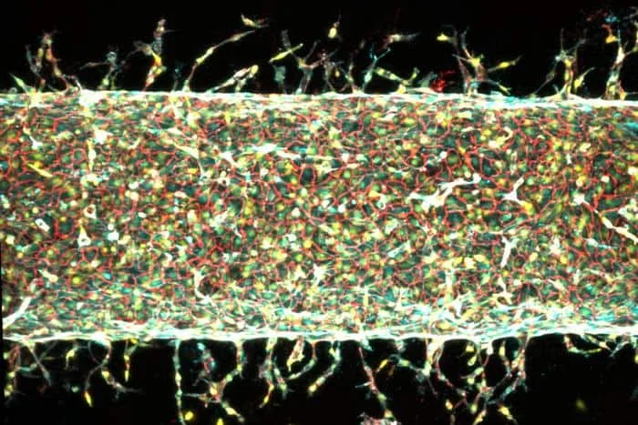
Tissue engineering offers a means to restore, maintain or replace biological tissues. But it is difficult to grow blood vessels using conventional fabrication techniques like 3D printing, as they are typically not precise enough to print fine capillaries and veins. A team of engineers from the Massachusetts Institute of Technology (MIT) has now developed a new approach to do just that, by using mechanical forces. Their blood-vessel-on-a-chip could be the first step in the scalable production of vascular networks that could then be implanted in the body to replace tissue that has been damaged by disease or injury.
The researchers, led by Ritu Raman, made their chip by filling a Petri dish with a gel containing nutrients and cell-growth factors. They then embedded a small magnet into the gel. Finally, they introduced a thin, hollow tube into the gel and coated the inside of the tube with live endothelial cells – the cells that naturally line blood vessels in the body.
Once anchored onto the inner walls of the tube, the endothelial cells began sprouting new, capillary-like vessels in the gel. And when the researchers moved the embedded magnet back and forth in different directions and by varying degrees, they found that the mechanical forces created by the moving magnet could stretch the vessels and increase the number of new capillaries that grew, as well as their length.
‘Steering’ blood vessel growth
“We also showed that blood vessel growth, or angiogenesis, could be directionally patterned using the mechanical forces created by the magnet and that these directions could change dynamically over time,” explains Raman. “This allows us to ‘steer’ blood vessel growth in all three spatial directions (x, y, z) and make complex geometries (like L-shaped branches) that would not be possible to fabricate using conventional methods.”
The researchers had previously demonstrated that biological cells are highly responsive to mechanical forces in the body, but they did not know how different types of forces – for example, stretching in different directions and at different degrees and frequencies — impacted the 3D structure of blood vessels. The main obstacle lay in developing a coupled magnet system to precisely stretch blood vessels in different directions. To achieve this, they designed a method for mechanically stimulating tissues, dubbed “magnetic matrix actuation”.
In their new work, which is detailed in PNAS, they went a step further and developed a method to accurately yet non-invasively stretch 3D blood vessel tissues they had built in the laboratory.
To better understand the effect of mechanical forces on angiogenesis, the MIT team decided to look into the role of a gene known as PIEZO1, which is just one of the many mechanically sensitive genes in the body. They built on the work of molecular biologist Ardem Patapoutian, who received the 2021 Nobel Prize in Physiology or Medicine for discovering that ion channels in cells open and close in response to mechanical pressure. One such ion channel is PIEZO1, which is regulated by the PIEZO1 gene.
Nanosilicates deliver angiogenesis therapies
By suppressing this PIEZO1 gene in the endothelial cells they studied, the researchers indeed observed that significantly fewer new blood vessels grew in the gel. This, they say, provides evidence that the applied mechanical forces in their experiment may have been opening the PIEZO1 ion channel in the endothelial cells, so triggering the new blood vessel growth.
“My group has always been interested in engineering artificial tissues that can be implanted in the body to repair damage caused by disease or traumatic injury, but such artificial tissues require integrated networks of blood vessels in order to function,” says Raman. “We hope that our method for precisely fabricating complex networks of blood vessels within engineered tissues will enable the development of such implants,” she tells Physics World.