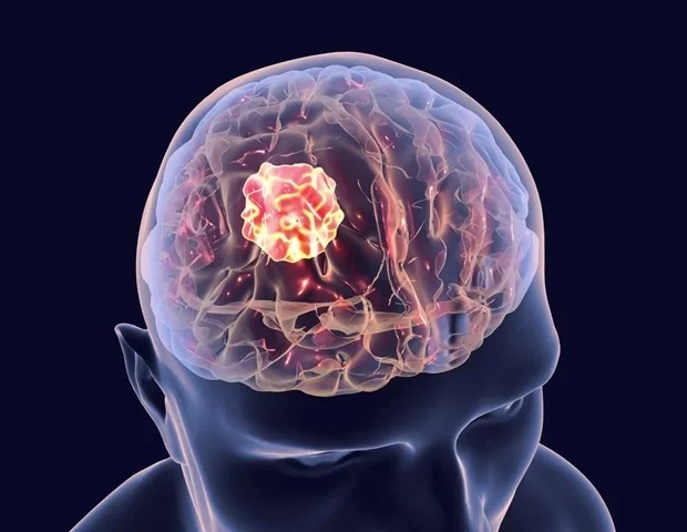
Glioblastoma is the most aggressive form of brain cancer.
It's difficult to treat because tumour cells infiltrate the surrounding brain tissue, making it hard for surgeons to remove the cancer completely without damaging healthy tissue. Treatment is complicated further by the blood-brain barrier, which limits how well drugs and radiotherapy reach the brain. All these factors contribute to a five-year survival rate of only around 7 per cent.
Researchers from the University of Technology Sydney (UTS), Harvard and Henan universities have discovered a 'double-punch' nanozyme platform designed to address both problems with a single tool of smart nanoparticles, published in Science Translational Medicine.
We've engineered a single material that does two jobs in sequence. It's a precise guide for the surgeon during the operation, and then a targeted clean-up treatment afterwards."
Dr. Bingyang Shi, Chair Professor of Nanomedicine, School of Electrical, Mechanical and Biomedical Engineering, University of Technology Sydney
The platform is built around an ultrathin, two-dimensional sheet studded with individually placed atoms, deposited one at a time using a technique borrowed from the semiconductor industry. This atomic-scale design gives the material two switchable functions: imaging during surgery and phototherapy after surgery, both activated by the same near-infrared light.
"During surgery, it functions as a highly sensitive imaging agent," said Professor Shi. "A fluorescent dye engineered onto the sheet glows under a near-infrared wavelength invisible to the naked eye, allowing surgeons to see individual tumour cell clusters as small as 44 micrometres, a resolution beyond current clinical imaging tools. A targeting molecule attached to the material also helps it cross the blood-brain barrier and accumulate specifically in glioma cells.
"After the visible tumour is removed, the same material is administered into the surgical cavity and reactivated with the same wavelength of light for postoperative phototherapy," he said. "The platinum atoms convert the tumour's own hydrogen peroxide into oxygen, counteracting the low-oxygen environment that normally shields cancer cells from treatment, while the light simultaneously generates heat and reactive molecules that destroy microscopic cancer cells that surgery could not reach."
In mouse models of glioblastoma the approach suppressed tumour recurrence after surgery, achieving 100 percent survival at 60 days in treated animals compared with a survival of 42 days for surgery alone, without inducing detectable neurological or motor deficits in follow-up testing.
"The results are very encouraging, but this is still early-stage research carried out in mouse models, not in people – and that distinction is important," said Professor Shi. "Its imaging and therapeutic performance will also need to be confirmed at the scale of a human brain.
"If this continues to hold up through that process, the hope is that surgeons could one day see more of the tumour during an operation and treat more of what's left behind afterwards. It's a meaningful step towards reducing recurrence, which remains one of the biggest challenges for people with glioblastoma."
Source:
Journal reference:
Shangguan, P., et al. (2026) Spatiotemporal-switchable 2D NIR-II single-atom nanozyme for single-cell–level surgical navigation and glioblastoma phototherapy. Science Translational Medicine. DOI:10.1126/scitranslmed.aeb8054. https://www.science.org/doi/10.1126/scitranslmed.aeb8054.